

An international research team has succeeded for the first time in using X-rays for an imaging technique that exploits a particular quantum property of light. The research team, led by Henry Chapman, leading scientist at DESY and professor at Universität Hamburg, used very intense X-ray pulses from the European XFEL to generate fluorescence from copper atoms. By measuring two photons from the emitted fluorescence almost simultaneously, scientists can obtain images of the copper atoms. The research, published in Physical Review Letters, could enable imaging of individual large molecules.
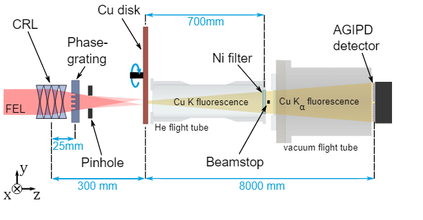
The atomic structures of materials and large molecules such as proteins are usually determined using X-ray crystallography, which relies on “coherent” X-ray scattering. Undesirable incoherent processes like fluorescence emission, however, can dominate the measurements, adding a featureless fog or background to the measured data. In the 1950s, astronomers Robert Hanbury Brown and Richard Twiss coined a method called “intensity interferometry”, that can extract structural information through the ‘incoherent’ fog. The method exploits the quantum mechanical properties of light, and opened the door to new understanding of light.
Read more on the European XFEL website
Image: The sum of over 58 million correlations of X-ray fluorescence snapshots is shown in the left insert, which was analysed by methods of coherent diffractive imaging to produce a high-resolution image of the source – here two illuminated spots in a spinning copper disk. Right insert: Reconstructed fluorescence emitter distribution at the copper disc with the two beam spots clearly visible.
Credit: DESY, Fabian Trost