New imaging technique for deeper insights in breast cancer metastasis

A collaborative effort between researchers from DESY, the University Medical Center Hamburg-Eppendorf (UKE), Chalmers University in Sweden and the Paul Scherrer Institute in Switzerland has yielded a cutting-edge multimodal imaging approach to investigate breast cancer tissue. With the help of this technique, researchers can simultaneously extract information about the nanostructure of the tumor and quantify the chemical elements present in a millimeter-scale sample in all three dimensions. A unique combination of research possibilities at PETRA III and new analysis methods enables this high level of detail. 

Breast cancer caused 685 000 deaths globally in 2020 according to the WHO. It is not life-threatening in its earliest form. But if the cancer cells are able to spread further in the tissue to nearby lymph nodes or important organs, this metastasis can be fatal. In a recent pilot study published in Nature Scientific Reports, the team applied this revolutionary imaging approach to a breast cancer sample. The results show how key molecules collectively influence the metastatic mechanism. This breakthrough paves the way for an in-depth investigation of breast cancer metastasis, promising novel therapeutic approaches and personalised treatment strategies, which could ultimately improve patients’ lives if recognized early enough.

Traditional experimental models often fall short, relying on 2D cell cultures or animal models that do not faithfully replicate the complex physiological patterns of human tumor environments. The multimodal imaging approach presented in this study represents a significant step forward by providing simultaneous nanoscale morphological and physiological information from real samples, thus giving researchers information about the shape and composition of real cancer tissue.

André Conceição, the first author and beamline scientist at the PETRA III SAXSMAT beamline P62, emphasises, “Although demonstrated for breast cancer, this approach’s versatility extends to other organs and diseases.”

The study opens avenues for further exploration of breast cancer metastasis and pre-metastatic niches (PMNs). Advanced X-ray multimodal tomography can generate complementary 3D maps for different breast cancer molecular subtypes. It holds the potential to contribute to the development of more targeted and effective strategies for diagnosis and treatment.

Read more on DESY website

Image: 3D vector field of the collagen direction and degree of orientation obtained by SAXS-Tensor-Tomography

Detection of early pancreatic cancer lesions using infrared and machine learning

A group of researchers from the CIRI beamline in their latest publication entitled Pancreatic intraepithelial neoplasia detection and duct pathology grading using FT-IR imaging and machine learning published in Spectrochimica Acta Part A: Molecular and Biomolecular Spectroscopy presented the results of their PanIN classification method, which provides opportunities for early recognition of changes in the cells lining the pancreatic ducts using infrared and machine learning.

Pancreatic intraepithelial neoplasia (PanIN) manifests itself by changes in the cells lining the pancreatic ducts. It is an early pre-cancerous lesion divided into low-grade and high-grade PanIN. In particular, high-grade PanIN is a lesion that often leads to Pancreatic ductal adenocarcinoma (PDAC). In the case of pancreatic cancer, due to the lack of characteristic symptoms of the disease in its early stage, patient survival is low. The basic examination performed to diagnose the disease is to take a fine-needle biopsy from the patient. The most common method of treatment is to remove part of the tissue affected by cancer, which increases the patient’s chances of survival, especially if it is done at an early stage of the disease. Therefore, it is so important to understand the biochemistry of lesions such as PanIN and their progression to cancer. 

Read more on SOLARIS website

Image : Scheme of sample collection (figure upper part), and FT-IR imaged TMA processing using: Random Forest classification (figure middle part), PLS Regression (figure bottom part).

Repairing genetic damage with sunlight

DNA damage to the genetic material DNA drives cancer, ageing, and cell death. Therefore, DNA repair is crucial for all organisms, and a deeper understanding of this basic function helps us better comprehend how life around us survives and thrives. An international team of researchers has now revealed how the enzyme photolyase efficiently channels the energy of sunlight into DNA repair chemistry.

All life under the sun must cope with harmful UV rays. UV damage can take many forms, but DNA, the molecule that carries the genetic information of all living organisms, is especially vulnerable. For instance, UV can drive chemical cross-linking reactions of DNA, potentially introducing errors into the genetic code. This cross-linking can lead to cell death or – in the worst cases – mutagenesis and cancer. Such damage is not uncommon; under bright sunlight, a human skin cell can undergo 50-100 cross linking reactions per second.

“To survive, life has evolved powerful DNA repair mechanisms. One especially elegant solution is provided by the enzyme photolyase,” explains DESY scientist Thomas J. Lane, who is also a researcher in the Cluster of Excellence “CUI: Advanced Imaging of Matter” at Universität Hamburg. The enzyme uses sunlight to repair damage caused by sunlight. Photolyase is able to recognize the location where UV irradiation has cross-linked DNA and grabs onto those bits of damaged DNA. Then, it can capture a blue photon from the sun, and use it to perform repair chemistry, turning the DNA back into its original, healthy form.

To better understand how photolyase works, the scientists were particularly interested first in the form of the enzyme immediately after absorbing a photon, but before repairing the DNA. Second, they wanted to find out the exact sequence of bond-breaking chemical reactions necessary to turn damaged DNA into healthy DNA. As a third step, the team sought to better understand how photolyase can specifically recognize which DNA is damaged.

Conducting time-resolved crystallography at the SwissFEL X-ray free-electron laser of PSI the scientists were able to capture the excited state of the photolyase chromophore, letting them understand how the enzyme efficiently channels the energy of sunlight into DNA repair chemistry. “This research was only made possible by the recent development of X-ray free-electron laser sources. Their intense femtosecond-duration pulses let us record flash X-ray photographs that freeze all atomic motion so that we can follow the reaction step by step at the speed of molecules,” says first author Nina-Eleni Christou from DESY.

Read more on PSI website

Image: PSI researcher Camila Bacellar is pleased about the success in precisely analysing the DNA repair enzyme photolyase at the Alvra beamline of the Swiss X-ray free-electron laser SwissFEL.

Credit: Paul Scherrer Institute/Markus Fischer

Bleomycin: cancer drug with a hidden flaw

Scientists at the B23 beamline of the Diamond Light Source have used synchrotron light to make an important discovery about a common cancer therapy. Bleomycin is used to shrink a variety of tumours, but little is known about how this drug interacts with proteins in the bloodstream. The beamline scientists used synchrotron-grade circular dichroism to study how bleomycin interacts with two common blood proteins, one of which is normally elevated in people with cancer. Reporting in the International Journal of Molecular Sciences, they found that the drug bound more firmly to the protein elevated in cancer patients, suggesting there may be less of the free form available to elicit its therapeutic effect. On closer analysis, the team discovered that one of two variants of bleomycin binds more strongly to this protein than the other. They caution that the ratio of these two variants may need to be adjusted to improve the therapeutic benefit of this drug.

While screening compounds produced by bacteria in the 1960s, scientists made a serendipitous discovery. They stumbled upon a molecule called bleomycin with anticancer properties. Since then, this life-saving drug has been used to treat a variety of tumours from squamous cell carcinomas to lymphomas. The drug works by chopping up DNA in cancer cells, and this DNA-drug interaction has been characterised in the past. However, when bleomycin enters the bloodstream, it may interact with plasma proteins and less is known about how this impacts the drug’s effectiveness. Bleomycin shows promising outcomes when tested on cancer cells grown in the lab, but the serum extracts used in lab cultures have a different mix of proteins to the sera of cancer patients, so it’s worth exploring whether plasma proteins in patients could sequester the drug and reduce its effectiveness.

Beamline scientists at B23 were determined to explore this overlooked issue. Led by Rohanah Hussain, they harnessed ultraviolet light from synchrotron radiation to explore how the drug binds plasma proteins using a technique called circular dichroism.

Circular dichroism is the differential absorption of left- and right-circularly polarised ultraviolet light passing through a liquid solution containing biomolecules, in this case proteins with drugs The CD measurement is displayed as a curve (spectrum) of which shape reflects the architecture on  how the protein in solution is folded in helical, ribbon, turn and unordered segments.  Drug binding to protein can affect such a folding that is used to identify and quantify drug binding interaction, in this case bleomycin with the two major blood proteins. Another unique experiment carried out at B23 beamline is the use of the powerful synchrotron beamlight to irradiate multiple of times the protein-drug mixtures for photostability assessment, which varies depending upon the strength of the drug binding interactions.

Hussain explained:

The high photon flux available at the B23 beamline (Diamond Light Source) generated by synchrotron radiation is sufficient for disrupting the folding of biological macromolecules in a time scale of minutes to hours, providing a useful tool for accelerated photo-stability studies.

First, the team assessed whether Blenoxane®, a commercial preparation of bleomycin, could bind to two common plasma proteins: one was human serum albumin (HSA), an abundant serum protein that facilitates the delivery of drugs around the body through the bloodstream. The other was α1-acid glycoprotein (AGP), a protein produced by the liver in response to inflammation that is found in cancer patients at ten times the normal level.

To explore binding interactions with these two proteins, the team examined the circular dichroism curve for each protein across a spectrum of ultraviolet light, and then they observed whether addition of Blenoxane® altered the protein curve. Sizeable differences were observed with AGP, suggesting the drug binds and induced marked changes to the protein’s shape, but the curve didn’t shift for HSA. This doesn’t indicate that the drug doesn’t bind HSA, only that it doesn’t alter its shape upon interaction. The team adapted their circular dichroism experiments to confirm that Blenoxane® did bind to HSA by heating the sample to unfold (denature) the proteins’ architecture and then observing spectral changes with and without the drug present.

Read more on Diamond Light Source  website

Image: Rohanah Hussain is a Senior Beamline Scientist, working on the B23 beamline at Diamond

Newly identified protein could help fight cancer

Researchers from the University of British Columbia (UBC) have identified a new protein that helps an oral bacterium thrive in other locations around the body. The discovery could eventually lead to the development of new drugs that specifically target the protein.

“This bacterium is common in the mouths of humans and generally doesn’t cause disease in that location. However, it can travel through the bloodstream to other areas of the body, which leads to some pretty big health concerns,” says Dr. Kirsten Wolthers, Associate Professor of Biochemistry and Microbiology at UBC’s Okanagan Campus.

Most notably, this bacteria is prevalent in the tumors of colorectal cancer patients. The presence of the bacteria can contribute to tumor growth, spread of cancer to other sites in the body, and resistance to chemotherapy.

With the help of the CMCF beamline at the Canadian Light Source (CLS), located at the University of Saskatchewan, Wolthers and her colleagues determined that the new protein they identified enables the bacteria to take essential nutrients, such as iron, from our blood cells.

Read more on the CLS website

Image: Alexis Gauvin, inspecting a protein sample for particulate matter, using the glove box. Gauvin is a biochemistry student and a member of Dr. Kirsten Wolthers’s research group in the Department of Chemistry, University of British Columbia (Okanagan Campus).

Tiny proteins found across the animal kingdom play a key role in cancer spread

Researchers from McGill University have made an exciting discovery about specific proteins involved in the spread of certain cancers.

Dr. Kalle Gehring, professor of biochemistry and founding director of the McGill Centre for Structural Biology, and his team have focused on unravelling the mystery around phosphatases of regenerating liver (PRLs). These proteins are found in all kinds of animals and insects — from humans to fruit flies – and play a unique role in the growth of cancerous tumours and the spread of cancer throughout the body.

“It’s important for us to study PRLs because they are so important in cancer,” said Gehring, “In some cancers, like metastatic colorectal cancer, the proteins are overexpressed up to 300-fold.”

This overexpression of PRLs makes cancer cells more metastatic and drives the spread to other organs.

In his most recent paper, published in the Journal of Biological Chemistry, Gehring and his colleagues confirmed that PRLs exist in all kinds of single- and multi-cell animals. Data collected at the Canadian Light Source (CLS) at the University of Saskatchewan confirmed the role of PRLs in binding magnesium transporters, helping to further the understanding of how these proteins influence human disease.

Read more on the Canadian Light Source website

Researchers study molecular bindings to develop better cancer treatments

A research team based in Winnipeg is using the Canadian Light Source (CLS) at the University of Saskatchewan to find new, cutting-edge ways to battle cancer.

Dr. Jörg Stetefeld, a professor of biochemistry and Tier-1 Canada Research Chair in Structural Biology and Biophysics at the University of Manitoba, is leading groundbreaking research into how netrin-1 — a commonly found molecule related to cell migration and differentiation —  creates filaments and binds to receptors in cells.

As netrin-1 is considered the key player for the migration of cancer cells, Stetefeld said this research could inform new cancer treatments.

“If you understand how netrin binds these receptors, you are sitting in the driver’s seat to develop approaches to block this interaction,” he said. “Why do we want to block it? Because if you block this interaction, you kill the cancer cell.”

Earlier research published in 2016 led to the development of new antibody treatments in Europe for combating breast cancer, said Stetefeld. He hopes this new research, which was published in the journal Nature, can lead to better drugs and treatments as well.

Read more on the CLS website

Using light to switch drugs on and off

Scientists at the Paul Scherrer Institute PSI have used the Swiss X-ray free-electron laser SwissFEL and the Swiss Light Source SLS to make a film that could give a decisive boost to developing a new type of drug. They made the advance in the field of so-called photopharmacology, a discipline that develops active substances which can be specifically activated or deactivated with the help of light. The study is being published today in the journal Nature Communications.

Photopharmacology is a new field of medicine that is predicted to have a great future. It could help to treat diseases such as cancer even more effectively than before. Photopharmacological drugs are fitted with a molecular photoswitch. The substance is activated by a pulse of light, but only once it has reached the region of the body where it is meant to act. And after it has done its job, it can be switched off again by another pulse of light.

This could limit potential side effects and reduce the development of drug resistance – to antibiotics, for example.

Licht-switchable drugs

To make conventional drugs sensitive to light, a switch is built into them. In their study, the scientists led by the principal authors Maximilian Wranik and Jörg Standfuss used the active molecule combretastatin A-4, which is currently being tested in clinical trials as an anti-cancer drug. It binds to a protein called tubulin, which forms the microtubules that make up the basic structure of the cells in the body, and also drive cell division. Combretastatin A-4, or “CA4” for short, destabilises these microtubules, thereby curbing the uncontrolled division of cancer cells, i.e. it slows down the growth of tumours.

In the modified CA4 molecule, a bridge consisting of two nitrogen atoms is added, which makes it particularly photoactive. In the inactive state, the so-called azo bridge stretches the molecular components to which it is attached to form an elongated chain. The pulse of light bends the bond, bringing the ends of the chain closer together – like a muscle contracting to bend a joint. Crucially, in its elongated form, the molecule does not fit inside the binding pockets of the tubulin – depressions on the surface of the protein where the molecule can dock in order to exert its effect. However, when the molecule is bent, it fits perfectly – like a key in a lock. Molecules like this, which fit into corresponding binding pockets, are also called ligands.

Read more on the PSI website

Image: Jörg Standfuss (left) and Maximilian Wranik in front of the experimental station Alvra of the Swiss X-ray free-electron laser SwissFEL, where the photopharmacological studies were carried out. In the long term, the aim is to develop drugs that can be switched on and off by light.

Credit: Paul Scherrer Institute/Markus Fischer

Antibody rigidity regulates immune activity

Scientists at the University of Southampton have gained unprecedented new insight into the key properties of an antibody needed to stimulate immune activity to fight off cancer, using the ESRF’s structural biology beamlines, among others.

The interdisciplinary study, published in Science Immunology, revealed how changing the flexibility of the antibody could stimulate a stronger immune response. The findings have enabled the team to design antibodies to activate important receptors on immune cells to “fire them up” and deliver more powerful anti-cancer effects. The researchers believe their findings could pave the way to improve antibody drugs that target cancer, as well as automimmune diseases.

In the study, the team investigated antibody drugs targeting the receptor CD40 for cancer treatment. Clinical development has been hampered by a lack of understanding of how to stimulate the receptors to the right level. The problem being that if antibodies are too active they can become toxic. Previous research by the same team had shown that a specific type of antibody called IgG2 is uniquely suited as a template for pharmaceutical intervention, since it is more active than other antibody types. However, the reason why it is more active had not been determined. What was known, however, is that the structure between the antibody arms, the so called hinges, changes over time.

This latest research harnesses this property of the hinge and explains how it works: the researchers call this process “disulfide-switching”. In their study, the team analysed the effect of modifying the hinge and used a combination of biological activity assays, structural biology, and computational chemistry to study how disulfide switching alters antibody structure and activity.

Read more on the ESRF website

Image: Flexibility of the monoclonal antibody F(ab) arms is conferred by the hinge region disulphide structure

Credit: C. Orr

Paving the way for more effective pancreatic cancer research

A team of scientists led by the University of Surrey used Diamond’s B16 Beamline, a flexible and versatile beamline for testing new developments in optics and detector technology and for trialling new experimental techniques, to better understand the structure of cancer cells. 

By using the synchrotron, the team were able to complete sophisticated examinations of the characteristics of cell structures at a nano level and even at an atomic scale and to investigate how cells and materials interact with each other.  

To improve cancer screening and treatment, researchers need accurate models of cancer tissues on which to experiment. Previous research made significant progress in building accurate, novel 3D models which mimic features of a pancreatic tumour, such as structure, porosity and protein composition.

Read more on the Diamond website

Image: Inside the experimental hutch at Diamond’s B16 beamline.

Credit: Diamond Light Source

Cell cytoskeleton as target for new active agents

Through a unique combination of computer simulations and laboratory experiments, researchers at the Paul Scherrer Institute PSI have discovered new binding sites for active agents – against cancer, for example – on a vital protein of the cell cytoskeleton. Eleven of the sites hadn’t been known before. The study is published in the journal Angewandte Chemie International Edition.

The protein tubulin is an essential building block of the so-called cell cytoskeleton. In cells, tubulin molecules arrange themselves into tube-like structures, the microtubule filaments. These give cells their shape, aid in transporting proteins and larger cellular components, and play a crucial role in cell division.

Thus tubulin performs diverse functions in the cell and in doing so interacts with numerous other substances. “Tubulin can bind an astonishing number of different proteins and small molecules, several hundred for sure,” says Tobias Mühlethaler, a doctoral candidate in the PSI Laboratory of Biomolecular Research and first author of the study. The functions of the protein are guided by means of such bonds. Also, many drugs dock on tubulin and take effect, for example, by preventing cell division in tumours.

Read more on the PSI website

Image: The research team in front of the Swiss Light Source (from left): Andrea Prota, Tobias Mühlethaler and Michel Steinmetz


Credit: Paul Scherrer Institute/Mahir Dzambegovic

How cellular proteins control cancer spread

New finding may help focus the search for anti-cancer drugs

A new insight into cell signals that control cancer growth and migration could help in the search for effective anti-cancer drugs. A team of researchers has revealed key biochemical processes that advance our understanding of colorectal cancer, the third most common cancer among Canadians.

Using the CMCF beamline at the Canadian Light Source (CLS) at the University of Saskatchewan, scientists from McGill University and Osaka University in Japan were able to unlock the behavior of an enzyme involved in the spread of cancer cells. The team found that there is a delicate interaction between the enzyme, PRL3, and another protein that moves magnesium in and out of cells. This interaction is crucial to colorectal cancer growth.

Read more on the Canadian Light Source website

Image: Members of the Gehring research laboratory discussing the results of a protein purification.

Promising new drug carrier could improve bone repair and cancer treatments

Researchers from Western University and the Shanghai Institute of Ceramics, Chinese Academy of Sciences used the Canadian Light Source (CLS) at the University of Saskatchewan to explore a promising drug carrier that could be used to deliver cancer treatments and therapeutics for severe injuries.

Their work advances drug carrier technology to make the carrier more compatible with our bodies. This allows the drug carrier to deliver the desired treatment precisely to a tumor, or to allow a slower release of the medicine. In a new paper published in The Royal Society of Chemistry, the team investigated using calcium phosphate as a potential drug carrier. Their approach uses phosphate from the biomolecule that stores and transports energy in our cells, which allows the carrier to be more compatible with the human body. Using this drug delivery system solves the limitations of other carriers, including biocompatibility and toxicity. Their carrier is highly compatible with our biological system, allowing for a better response while also being non-toxic.

“Calcium phosphate is an important biomaterial in bones and teeth. If you can use this material as a drug carrier then you don’t need to worry about what happens after it is done with delivery,” said Tsun-Kong (TK) Sham, Professor of Chemistry at Western University.

Read more on the Canadian Light Source website

Image: TK Sham, a Professor of Chemistry at Western University, using beamlines at the CLS.

Faster diagnosis of esophageal cancer

Scientists from SOLARIS National Synchrotron Radiation Centre (Kraków, Poland), University of Exeter (UK), Beckman Institute (University of Illinois at Urbana-Champaign – USA), and Institute of Nuclear Physics Polish Academy of Sciences (Kraków, Poland) performed research that will facilitate the rapid and automated diagnosis of esophageal cancer.

Dr. Tomasz Wróbel`s group focuses on cancer detection through a combination of Infrared Imaging (IR) and the use of Machine Learning (ML) algorithms. Thanks to this approach, it is possible to develop an effective model, which will allow histopathologists to confirm the diseased area automatically and in a much shorter time.

Infrared Imaging (IR), which will soon also be available on the newly built beamline at SOLARIS, has found widespread use in biomedical research over the last couple of decades and is currently being introduced into clinical diagnostics.

Read more on the Solaris website

Image: The above graphic shows two esophageal biopsies: the top of the graphic contains a biopsy taken from a patient suffering from esophageal cancer, the bottom of the graphic contains a biopsy taken from a healthy patient. In the left part of the graphic, microscopic images of the mentioned biopsies are visible after the H&E staining (Hematoxylin and Eosin) (in this image of the stained biopsy, the histopathologist visually assigns tissue types), in the middle of the graphic, biopsy images obtained using infrared imaging are visible, the right part of the graphic presents a histological picture of a biopsy obtained after assigning tissues and structures to three classes (cancer, other, benign) by Machine Learning (ML).

Red – cancer
Blue – other
Green – benign

Developing microbeam radiation therapy for inoperable cancer

An innovative radiation treatment that could one day be a valuable addition to conventional radiation therapy for inoperable brain and spinal tumors is a step closer, thanks to new research led by University of Saskatchewan (USask) researchers at the Canadian Light Source (CLS).

Microbeam radiation therapy (MRT) uses very high dose, synchrotron-generated X-ray beams—narrower than a human hair—to blast tumours with radiation while sparing healthy tissue. The idea is that MRT would deliver an additional dose of radiation to a tumor after maximum conventional radiation therapy has been tried, thereby providing patients with another treatment that could extend their lives. 

But the longstanding questions have been: What is the optimal X-ray energy range of the MRT radiation dose that will both penetrate the thickness of the human body and still spare the healthy cells? How can the extremely high radiation doses be delivered and measured with the accuracy necessary for human treatment?

Read more on the Canadian Light Source website

Image : Farley Chicilo at the Canadian Light Source.

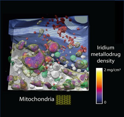
Imaging how anticancer compounds move inside the cells

Chemotherapeutics are key players in the clinical setting to fight most types of cancer, and novel chemicals hold the promise to facilitate new and unique intracellular interactions that modulate the cell machinery and destroy the tumour cells. Equally necessary are new tools that enable the unequivocal location and quantification of such molecules in the intracellular nano-space, so that their therapeutic action is fully understood.

Researchers from IMDEA Nanociencia, the ALBA Synchrotron, the European Synchrotron Radiation Facility (ESRF) and the National Centre for Biotechnology (CNB) have developed a new family of organo-iridium drug candidates about a hundred times more potent than the clinically used drug cisplatin.
In order to understand the therapeutic potential of the compound, it is mandatory to accurately localize its fate within the cell ultrastructure with minimal perturbation. To this aim researchers have correlated on the same cell, for the first time, two 3D X-ray imaging techniques with a resolution of tenths of nanometers: cryo soft X-ray tomography, at MISTRAL beamline at ALBA Synchrotron, and cryo X-ray fluorescence tomography, at ID16A beamline at ESRF. These techniques help elucidate the 3D architecture of the whole cell and to reveal the intracellular location of different atomic elements, respectively.

>Read more on the ALBA website