

Successful tests confirm outstanding performance of coils
Researchers at European XFEL have reached an important milestone in developing a new generation of X-ray light sources. A set of superconducting electromagnets, produced by Bilfinger Nuclear, have proven their excellent performance, paving the way for the use of the design in future superconducting undulators. These devices will cause accelerated electrons to radiate much more effectively than current state-of-the-art technology allows. European XFEL aims to become the world’s first X-ray free-electron laser to use superconducting undulators. These undulators will unlock new research in fields such as materials science, chemistry, biology and high-energy-density science by providing X-ray pulses with significantly shorter wavelengths than have been possible at XFELs to date.
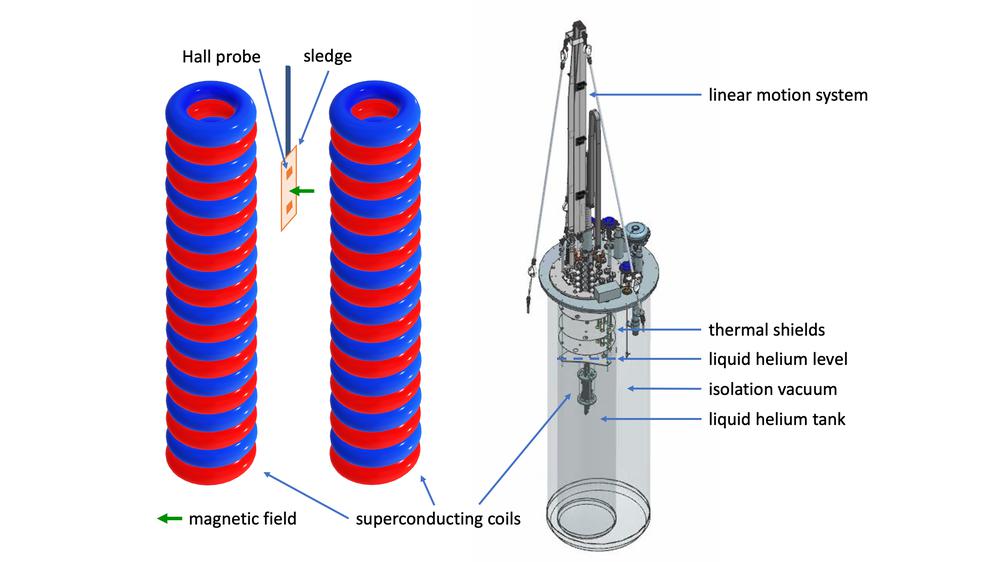
The magnetic field of undulators is designed to be highly periodic, precisely controlled, and exceptionally uniform along the electron beam path. The electromagnets examined at European XFEL consist of niobium-titanium (NbTi) wire. At the operating temperature of -269 degrees Celsius, the material is superconducting, meaning it can carry very high electrical currents with virtually no resistance. When wound into coils with extreme precision, an electromagnet is created that produces a strong magnetic field when carrying an electric current. Measurements of their magnetic field have now been completed and show that the coils successfully reached the required operating current and produced the target magnetic field of 1.82 Tesla, while maintaining the necessary field quality for X-ray generation over the entire 2-metre length of the coils.
This is important because using the devices to generate X-rays relies not only on the magnetic fields being very strong, but also on them being highly periodic. Even tiny deviations from this periodic structure affect the quality of the X-ray beam generated. The qualification tests demonstrate that the coils can meet these demanding requirements over their full two-metre length, making them the longest high-precision superconducting undulator coils ever produced and measured.
Read more on the European XFEL website
Image: The SUNDAE1 test stand and a sketch of the sledge attached to a rod with Hall probes sliding along the magnetic field axis of the SCU coils (Illustration: S. Casalbuoni et al., Front. Phys. Sec. Interdisciplinary Physics Volume 11 – 2023)


