
Chemotherapeutics are key players in the clinical setting to fight most types of cancer, and novel chemicals hold the promise to facilitate new and unique intracellular interactions that modulate the cell machinery and destroy the tumour cells. Equally necessary are new tools that enable the unequivocal location and quantification of such molecules in the intracellular nano-space, so that their therapeutic action is fully understood.
Researchers from IMDEA Nanociencia, the ALBA Synchrotron, the European Synchrotron Radiation Facility (ESRF) and the National Centre for Biotechnology (CNB) have developed a new family of organo-iridium drug candidates about a hundred times more potent than the clinically used drug cisplatin.
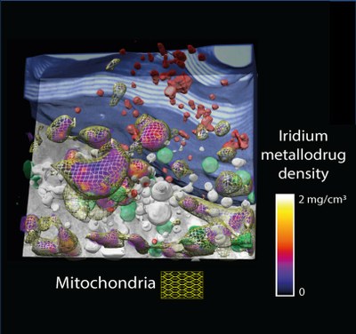
In order to understand the therapeutic potential of the compound, it is mandatory to accurately localize its fate within the cell ultrastructure with minimal perturbation. To this aim researchers have correlated on the same cell, for the first time, two 3D X-ray imaging techniques with a resolution of tenths of nanometers: cryo soft X-ray tomography, at MISTRAL beamline at ALBA Synchrotron, and cryo X-ray fluorescence tomography, at ID16A beamline at ESRF. These techniques help elucidate the 3D architecture of the whole cell and to reveal the intracellular location of different atomic elements, respectively.
>Read more on the ALBA website