

Widely used models mispredict collective electron oscillations in warm dense aluminium, study shows
Researchers at European XFEL, Helmholtz-Zentrum Dresden-Rossendorf (HZDR), Rostock University and other collaborating institutions have used high-precision experiments to demonstrate that the most widely used models for the behaviour of electrons in warm dense matter are inaccurate. Warm dense matter is challenging to study but also of key importance for a plethora of research, including the investigation of planetary interiors, material science, and laser fusion experiments. The study has been published in Physical Review Letters.
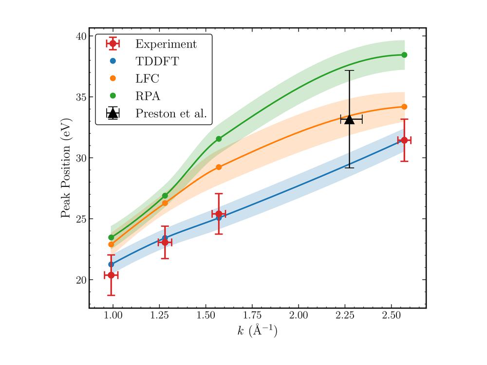
In warm dense matter, electron density oscillates. The collective oscillations are called plasmons. They carry important information and can be observed using X-rays, resulting in scattering spectra – abstract images captured by a detector. In many experiments, these spectra are interpreted using simplified uniform electron gas models. However, the new measurements show that, for warm dense aluminium, these models consistently overestimate the plasmon energy by up to about 25 per cent (about 8 electronvolts) and fail to reproduce the full measured shape of the signal.
“Our measurements are precise enough to clearly distinguish between competing models,” says Thomas Preston of European XFEL. “That is important because these models are widely used to diagnose extreme states of matter. If the model is incorrect, that leads to inaccurately inferred properties.” The electron behaviour affects predictions of opacity, optical properties, electrical conductivity, and energy transport, for instance.
Read more on the European XFEL website
Image: Experimental setup at the HED-HIBEF instrument
Credit: European XFEL

