European XFEL elicits secrets from an important nanogel

An international team led by Felix Lehmkühler from Deutsches Elektronen-Synchrotron DESY in Hamburg has investigated the temperature induced swelling and collapsing of the polymer poly-N-isopropylacrylamide (PNIPAm) at European XFEL at Schenefeld near Hamburg. Due to its dynamic changes, PNIPAm is used in medicine, e.g. for drug delivery, tissue engineering or sensorics.

PNIPAm is typically dissolved in water. Above a certain temperature, the so-called lower critical solution temperature (LCST), which is around 32 °C, it changes from a hydrophilic, water-loving state to a hydrophobic, water-repellent state. As consequence, nanogel particles, as investigated by Lehmkühler and co-workers, rapidly change their size above that temperature by expelling water.

This feature is useful for a variety of applications, including the controlled release of drugs in a patient’s body, as a model system for proteins and in tissue engineering, the cultivation of organic tissue for medical applications, or as bio-compatible temperature sensors. However, it was very difficult so far to watch these rapid phase transitions experimentally, and therefore to optimize them for different applications. Therefore, the precise characterisation of the kinetics of the changes of the PNIPAm polymer with temperature is still a lively research topic.

Read more on XFEL website

Image: Felix Lehmkühler at the instrument MID (Materials Imaging & Dynamics) of European XFEL where the experiments were done.

Credit: European XFEL

Röntgen medal for Robert Feidenhans’l

The Danish physicist Robert Feidenhans’l has spent almost his entire career working in the field of X-ray synchrotron radiation and free-electron lasers and is regarded as a pioneer in the use of X-rays from synchrotron radiation facilities. He is a co-founder of surface crystallography and was the first person to succeed in precisely determining surface structures experimentally. Feidenhans’l is an outstanding researcher who always regarded science as a means of promoting international understanding and collaboration.

His research includes nanophysics, in particular research into nanowires and the development of X-ray techniques for analysing materials. He was also instrumental in the development of X-ray imaging methods for the three-dimensional characterisation of materials and biological tissue, and was involved in high-resolution micro X-ray tomography in medicine, for example to investigate the interaction between bones and implants.

Between 2017 and 2023, Feidenhans’l was Chairman of the Management Board of the European X-ray laser and responsible for the transition from construction into full user operation. The international research facility in Schenefeld near Hamburg is one of the most powerful X-ray lasers in the world.

During his career, he has held numerous positions in science management. For example, he was Chairman of the Danish National Committee for Crystallography (1998-2007), Chairman of the Council of the European Synchrotron Radiation Facility (ESRF) (2006-2010), Chairmen of the European XFEL Council (2010-2014), Member of the Danish Academy of Technical Sciences (ATV) (since 2015), Chairman of the Board of MAX IV (since January 2023), member of the Board of Trustees of the Joachim Herz Foundation (since January 2023) and Member of the Scientific Advisory Board at the Advanced Light Source in Berkeley, USA (since 2024).

Read more on XFEL website

Image: The German Röntgen Museum honours Robert for his work on X-ray imaging methods.

Credit: European XFEL

Milestone for superconducting undulator

A EUROPEAN XFEL TEAM AT THE KARLSRUHE INSTITUTE FOR TECHNOLOGY HAS TESTED A MOCK-UP COIL OF THE SUPERCONDUCTING UNDULATOR PRE-SERIES MODULE (S-PRESSO) DESIGNED FOR AN UPGRADE OF THE EUROPEAN XFEL. IT REACHED A WORLD RECORD MAGNETIC FIELD.

Undulators are one of the most important devices for a free-electron laser like the European XFEL in Schenefeld near Hamburg. With the help of a series of strong magnets an undulator creates an extremely brilliant light by forcing fast-moving electrons onto a slalom course. Furthermore, the undulators stimulate the electrons to emit laser-like electromagnetic radiation.

The strength of the magnets of an undulator determines the tunability of the photon energy range available for experiments. The Undulator Systems Group of European XFEL has started different activities in collaboration with Deutsches Elektronen-Synchrotron DESY to allow the implementation of superconducting undulators into the European XFEL in the upcoming years. The contract for superconducting undulator pre-series module (S-PRESSO) consisting of two pair of coils and a phase shifter has been assigned to Bilfinger Noell GmbH. Now, a European XFEL team at the Karlsruhe Institute for Technology has tested a 30-centimeter-long mock-up superconducting coil designed and build by Bilfinger Noell GmbH. The magnetic field of the S-PRESSO mock-up has reached 2 Tesla, which is larger ever reached before in such undulators.

Read more on the European XFEL website

Image: Undulators like this one cause highly accelerated electrons to emit intense and brilliant X-ray light. European XFEL is currently testing superconducting undulators in order to be able to offer users even better conditions for their research in the future.

European XFEL Young Scientist Award for Jiawei Yan

Dr. Jiawei Yan was awarded the 2,000-euro prize for young scientists for the development of methods for generating ultrashort high-power pulses at the SASE2 line of the European XFEL.

He and a team from the FEL Physics group of the European XFEL and the MXL group at DESY, which is responsible for the operation of the XFEL, developed a method to achieve remarkably sharp current profiles with high peak currents.

Read more on XFEL website

Image: Marc Simon, Co-Chair of the User Organization Executive Committee, ceremonially presents the award to Jiawei Yan. 

Credit: European XFEL

Transition metal insulators: The origin of colour

In a theoretical study, researchers have explained the vibrant colours of two compounds whose electronic properties seemingly prohibit such colouring. The hues exhibited by the two insulators originate from transitions in the spins of the electrons, which modify the way the materials absorb and reflect light in such a way as to create the bright colours. The theoretical framework employed by the team promises new insights in fields such as optoelectronics or in the study of qubits, the quantum bits used in quantum computers. 

Although colour is a familiar phenomenon, it is sometimes challenging to explain how the hues of certain materials come about. This is the case with insulators that contain transition metals. In these compounds, the energy gap between the valence band, in which the electrons are tightly bound to the atoms, and the conduction band, in which the electrons can move freely, is larger than the highest energy of photons of visible light—meaning that these materials should not absorb visible light. As the colour of a compound is complementary to the wavelengths it absorbs, we should thus perceive these insulators as being transparent instead of coloured. 

A team of researchers including the head of the European XFEL Theory group, Alexander Lichtenstein, now used two complementary theoretical methods to study the origin of colour in two typical transition metal insulators: nickel(II) oxide (NiO)—a green compound used in the production of ceramics and nickel steel as well as in thin-film solar cells, nickel–iron batteries, and fuel cells—and manganese(II) fluoride (MnF2), a pink material employed in the manufacture of special kinds of glass and lasers.

Read more on XFEL website

Image: Visualization of the orbital character of low-laying excitons in NiO, corresponding to a local ‘Frenkel’ exciton at an energy of 1.6 eV and a weakly bound, bright ‘Wannier-Mottâ’ exciton at an energy of 3.6 eV

Milestone for novel atomic clock

X-ray laser shows possible route to substantially increased precision time measurement

An international research team has taken a decisive step toward a new generation of atomic clocks. At the European XFEL X-ray laser, the researchers have created a much more precise pulse generator based on the element scandium, which enables an accuracy of one second in 300 billion years – that is about a thousand times more precise than the current standard atomic clock based on caesium. The team presents its success in the journal Nature.

Atomic clocks are currently the world’s most accurate timekeepers. These clocks have used electrons in the atomic shell of chemical elements, such as caesium, as a pulse generator in order to define the time. These electrons can be raised to a higher energy level with microwaves of a known frequency. In the process, they absorb the microwave radiation. An atomic clock shines microwaves at caesium atoms and regulates the frequency of the radiation such that the absorption of the microwaves is maximised; experts call this a resonance. The quartz oscillator that generates the microwaves can be kept so stable with the help of resonance that caesium clocks will be accurate to within one second within 300 million years.

Crucial to the accuracy of an atomic clock is the width of the resonance used. Current caesium atomic clocks already use a very narrow resonance; strontium atomic clocks achieve a higher accuracy with only one second in 15 billion years. Further improvement is practically impossible to achieve with this method of electron excitation. Therefore, teams around the world have been working for several years on the concept of a “nuclear” clock, which uses transitions in the atomic nucleus as the pulse generator rather than in the atomic shell. Nuclear resonances are much more acute than the resonances of electrons in the atomic shell, but also much harder to excite.

At the European XFEL the team could now excite a promising transition in the nucleus of the element scandium, which is readily available as a high-purity metal foil or as the compound scandium dioxide This resonance requires X-rays with an energy of 12.4 kiloelectronvolts (keV, which is about 10,000 times the energy of visible light) and has a width of only 1.4 femtoelectronvolts (feV). This is 1.4 quadrillionths of an electronvolt, which is only about one tenth of a trillionth of the excitation energy (10-19). This makes an accuracy of 1:10,000,000,000,000 possible. “This corresponds to one second in 300 billion years,” says DESY researcher Ralf Röhlsberger, who works at the Helmholtz Institute Jena, a joint facility of the GSI Helmholtz Centre for Heavy Ion Research, the Helmholtz Zentrum Dresden-Rossendorf (HZDR), and DESY.

Read more on the DESY website

Image: An artist’s rendition of the scandium nuclear clock: scientists used the X-ray pulses of the European XFEL to excite in the atomic nucleus of scandium the sort of processes that can generate a clock signal – at an unprecedented precision of one second in 300 billion years.

Credit: European XFEL/Helmholtz Institute Jena, Tobias Wüstefeld/Ralf Röhlsberger

Molecular motion in tire rubber

Scientists have observed the molecular motion of rubber components typically used in automobile tires—polybutadiene and carbon black—with the world’s fastest time resolution. The study reveals a clear interaction between the two components on the atomic scale, paving the way towards improved diagnostics of tire rubber degradation and the development of materials with enhanced durability.

Tire rubber is a composite material that typically includes synthetic rubber, such as polybutadiene, and added nanoparticles, such as carbon black, to improve its physical properties. During driving, strong forces act on the tire, causing its components to move against each another, which can lead to wear and degradation of the material. To evaluate tire performance, it is therefore important to understand not only the static structure of the complex particle network formed by the polymer and the nanoparticles, but also their interaction and respective movements, as these dynamics directly influence material properties such as wear resistance. Because some of these molecular movements happen extremely quickly, time-resolved measurements at atomic resolution on the fastest possible time scale are critical for developing and validating dynamic models of such materials.  

An international research team led by scientists from the University of Tokyo, Ibaraki University, and European XFEL has now observed the molecular motion within samples of polybutadiene and carbon black, which occurs naturally as a result of the material structure, with a time resolution of 890 nanoseconds (billionths of a second)—the fastest resolution obtained in such studies so far—at the European XFEL’s SPB/SFX instrument. 

Read more on XFEL website

Helium droplets for studies of nanostructures

Using a conical nozzle, an international research team has generated vortex-free droplets of superfluid helium that are larger than any created before. The droplets are big enough to be resolved in X-ray diffraction images, making them ideal for studying the self-assembly of a wide range of nanostructures forming inside a superfluid environment.  

A superfluid, such as very cold liquefied helium, flows without any internal friction. Droplets of superfluid helium therefore provide a perfect environment for researchers to investigate the formation of self-organized nanostructures made from various dopant materials, i.e. atoms and molecules specifically inserted into the droplets. However, the occurrence of vortices inside the droplets can hinder the assembly of such nanostructures, as many dopants are easily attracted to them. Now, a team of scientists led by researchers from TU Berlin has used a special nozzle at the European XFEL’s SQS instrument to create swirl-free helium nanodroplets and explore the size range in which they can be produced.

“Our conical nozzle enabled us to generate vortex-free droplets from the condensation of expanding helium gas that contain up to a thousand times more helium atoms than possible with previous methods,” explains Rico Tanyag, previously at TU Berlin in Germany and now at Aarhus University in Denmark, one of the principal investigators of the experiment. “This large size allows us to image both the droplets and the dopant nanostructures inside them using the ultrashort pulses of X-ray free-electron lasers such as the European XFEL,” adds Daniela Rupp from ETH Zürich in Switzerland, the other main proposer of the study. “Our experiment thus paves the way for exploring in atomic detail how such nanostructures form.”

Using a technique called X-ray coherent diffractive imaging on helium droplets doped with xenon atoms, the scientists found that single compact xenon structures, which are associated with vortex-free formation, prevailed up to a droplet size of a hundred million helium atoms—a thousand times more than previously feasible. Larger droplets, on the other hand, contained xenon filaments, indicating the presence of vortices that disturbed the structure formation. 

Read more on XFEL website

Image: The SQS instrument at European XFEL.

Credit: European XFEL/Axel Heimken

European XFEL control software Karabo released as open source

European XFEL has released the control software framework Karabo and selected Karabo devices into the public domain as free and open-source software, enabling external developers to use and adapt the code as they need. The extendable system can be used to control installations that range from single machines to highly complex research facilities, such as the European XFEL.

Since its first inception in 2011, the European XFEL control system has been developed into a modern, distributed software framework that enables control and monitoring of the photon systems and instrumentation at the facility, as well as data acquisition from X-ray detectors capable of megahertz frame rates. It is highly interoperable with DOOCS, a similar system developed at DESY that is used to control the European XFEL accelerator.

“Karabo is a distributed system which, thanks to the use of a modern message broker, can run on a single computer or on hundreds of servers, and which has grown with the facility,” emphasizes Dennis Goeries from the Controls group, who leads the development team responsible for the framework. “It is also highly scalable—it can run on a small system on a chip such as a Raspberry PI or on a system with tens of computing cores.”

“Functionality can be added to the core system through so-called devices,” explains Wajid Ehsan, head of the team developing the devices that are used throughout the European XFEL facility. “This enables Karabo developers to integrate hardware, define high-level procedures, and implement data acquisition, so they can tailor the system precisely to the facility they want to control.” At the European XFEL, the Karabo control system handles about 3.7 million control parameters on 30 000 devices over 13 major installations.

Read more on the European XFEL website

Image: Current and former Karabo contributors at the open-source release event.

Credit: European XFEL/Kai-Erik Ballak

New technique reveals insights on Vitamin B12

Researchers have implemented a new technique, based on the European XFEL’s ultrashort pulses, to gain insights into two compounds featuring vitamin B12. B12 is an important compound in many biological systems, and the new method will allow scientists to develop a much deeper understanding of its structure and behavior. The technique will enable better insights for a host of biological molecules, and could help in designing targeted drug therapies.

Scientists used the European XFEL’s bright and ultrashort X-ray pulses to probe the evolution of two B12 compounds in time, at intervals of just 10 trillionths of a second (100 fs). Scientists used green light to illuminate the B12 compounds, and looked at them using a new method of X-ray spectroscopy called time-resolved Valence-to-Core X-ray Emission Spectroscopy (tr-VtC XES) to take snapshots of each molecule focusing on different aspects of their structure. Through the combination of optical and X-ray measurements, researchers were able to learn about the specific behaviors of these B12 compounds, such as their reactions to visible light, as well as the way the molecules vibrate and recover their initial configuration.

“Similar techniques have been used to investigate B12 using only visible and ultraviolet light,” says Frederico Lima, Instrument Scientist at the FXE instrument. “But the advantage of using tr-VtC XES is that you can get an element sensitive measurement that is also rather straightforward to predict using modern quantum chemical theory. In other words, it becomes easier to understand what each individual element in the molecule is doing. This gives us a more precise picture of the vitamin’s behavior than previously possible.”

The study, published in the Journal of the American Chemical Society, also addresses a problem with measuring tr-VtC XES on biological systems such as those containing B12, namely, that they produce small, difficult to detect signals.

Read more on the European XFEL website

Image: Visualisations of the structure of two vitamin B12 compounds at a 2.6 angstrom resolution, (a) in reaction with a biological structure and (b) in a water-based environment.

New imaging technique could shed light on individual molecules


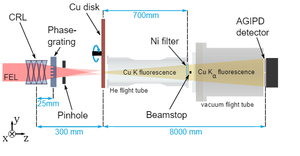
An international research team has succeeded for the first time in using X-rays for an imaging technique that exploits a particular quantum property of light. The research team, led by Henry Chapman, leading scientist at DESY and professor at Universität Hamburg, used very intense X-ray pulses from the European XFEL to generate fluorescence from copper atoms. By measuring two photons from the emitted fluorescence almost simultaneously, scientists can obtain images of the copper atoms. The research, published in Physical Review Letters, could enable imaging of individual large molecules.

The atomic structures of materials and large molecules such as proteins are usually determined using X-ray crystallography, which relies on “coherent” X-ray scattering. Undesirable incoherent processes like fluorescence emission, however, can dominate the measurements, adding a featureless fog or background to the measured data. In the 1950s, astronomers Robert Hanbury Brown and Richard Twiss coined a method called “intensity interferometry”, that can extract structural information through the ‘incoherent’ fog. The method exploits the quantum mechanical properties of light, and opened the door to new understanding of light.

Read more on the European XFEL website

Image: The sum of over 58 million correlations of X-ray fluorescence snapshots is shown in the left insert, which was analysed by methods of coherent diffractive imaging to produce a high-resolution image of the source – here two illuminated spots in a spinning copper disk. Right insert: Reconstructed fluorescence emitter distribution at the copper disc with the two beam spots clearly visible.

Credit: DESY, Fabian Trost

Unveiling finer details in the physics of materials

Scientists at the European XFEL’s SCS instrument routinely use a technique called transient X-ray absorption spectroscopy (XAS) to investigate materials that have applications in data storage and processing, catalysis, or in the search for room temperature superconductors. Investigating very small changes in the motion of electrons within a material’s structure on ultrashort timescales provides scientists with fingerprints of the complex processes at play within them. This helps them characterise samples that are important for energy and materials research.

Using the European XFEL’s brilliant pulses, researchers can overcome some of the issues of conventional transient XAS—such as long measurement times—but the varying intensity of European XFEL’s pulses provides its own challenges. Now, scientists at SCS have implemented a new sampling scheme for improving the efficiency of such measurements.

Read more on the European XFEL website

Image: The X-ray beam is split into three copies. Two of these copies are passed through identical samples of the material under investigation, with one of these samples also being illuminated by a laser (‘optical laser’ in the figure). This transforms it into a new state, interesting to researchers. From this, scientists are able to ‘subtract’ detrimental noise, revealing the finest details of the sample under investigation.

2023 Young Scientist Award winner announced

Dr Elke de Zitter, from the Institut de Biologie Structurale (IBS) in Grenoble, is the winner of the European XFEL Young Scientist Award 2023. The price was awarded today at the Users’ Meeting 2023 by Andrea Eschenlohr, chairwomen of the European XFEL User Organisation Executive Committee. De Zitter’s research focuses on processing serial-crystallography data taken by the SPB/SFX instrument at European XFEL. She is interested in mosquitocidal proteins, that target and kill mosquitos, the deadliest animal on Earth because of the diseases they carry. She has also worked on developing a piece of software known as Xtrapol8 which can extract protein structures from European XFEL data.

“The European XFEL Young Scientist Award highlights the future potential of young scientists working in X-ray laser science, outlining the talent, hard-work and dedication of the early-career researchers within our user community,” says Sakura Pascarelli, Scientific Director at European XFEL. “It is an opportunity to highlight the impact of new research done by talented young researchers, as well as to showcase the large collaborative efforts that are required for research at European XFEL.” 

Read more on the European XFEL website

New simulation tool opens path to superfast electronic switches

Electronic devices operate at speeds limited by the physical processes underlying their operation: the faster the process, the quicker the information processing speed. One such fast process that might lead to the development of superfast magnetic switches is the demagnetisation of layered magnetic materials (multilayered ferromagnets) when hit by ultrafast X-ray laser pulses. This process has been poorly understood to date, but now a joint research project by European XFEL and the Institute of Nuclear Physics of the Polish Academy of Sciences (IFJ PAN) has developed a new simulation tool, taking an important step towards superfast electronics.

“In recent years, physicists have become quite familiar with demagnetisation processes initiated by visible or near-infrared light. However, when it comes to the impact of more energetic X-ray radiation, we are all just taking the first steps,” says Beata Ziaja-Motyka, initiator of the research project. “Our team’s contribution lies in the construction of a theoretical model called XSPIN. With its help, it is possible for the first time to simulate demagnetisation in multilayered ferromagnetic materials exposed to femtosecond pulses of light from an X-ray laser.”

Read more on the European XFEL website

Image: A pulse of X-ray radiation hits a sample of material with magnetic properties, scatters and forms a diffraction ring. The diameter of the ring depends on the average distance between the magnetic domains, and its intensity is the greater, the stronger the magnetization of the sample.

Credit: FJ PAN

Ultrafast surface processes observed

SINGLE X-RAY LASER PULSES CAN BE USED TO OBSERVE ULTRAFAST CHANGES ON MATERIAL SURFACES WITH UNPRECEDENTED DEPTH AND TIME RESOLUTION

In a world first, an international team of scientists led by European XFEL and the University of Siegen has demonstrated that the intense pulses produced by an X-ray laser can be used to investigate ultrafast processes occurring on and just below material surfaces with unprecedented depth and time resolution. This allows researchers to capture processes that are more than a billion times faster than what could previously be observed. The results, which the team has just published in Physical Review Research, pave the way for versatile applications that rely on our understanding of ultrafast surface dynamics. Examples are the laser processing of material surfaces to create tailor-made nanoscale structures or the realization of compact laser-based particle or radiation sources.

Using intense laser pulses, nanoscale surface structures can be created with optimized optical, mechanical, and chemical properties. Such tailored structures play a decisive role in many fields with significant societal and economic impact. They can be used to fashion antimicrobial coatings, to improve the bonding of dental implant screws with bone, and to build advanced optical components with high damage thresholds. To be able to better create these structures and comprehend their effects, scientists first need to observe and understand the ultrafast processes that happen when the intense femtosecond laser pulses used in the surface processing hit the material and react with it.

Read more on the European XFEL website

Image: Grazing-incidence small-angle X-ray scattering image obtained from a multilayer sample, measured using single X-ray pulses of the SACLA X-ray laser in Japan. The central black circle is the beamstop used to block the main mirror-like reflection peak, which is much more intense than the scattering pattern. The pattern contains information on the depth-resolved density profile (horizontal axis) and the surface structure (vertical axis).

Karen Appel’s #My1stLight

Karen was a beamline scientist at DESY and is currently a beamline scientist at the European XFEL

My first synchrotron experiment was at beamline L at DORIS at DESY, which at that time just set up the possibility to do micro-focus X-ray fluorescence measurements. The first experiment I was involved in was headed by the group of Prof Schenk at the Institute of Mineralogy of the University of Kiel  and focused on minerals that were formed at high pressures and high temperatures. At that moment, I was a PhD student at the University of Bonn, working on metamorphic rocks and isotope geochemistry of rocks and got involved in the experiment, because I was interested in analytical methods that could be applied to minerals that were formed at high pressures and temperatures. Besides some connections through my earlier studies, my main interest was to learn about this new method of X-ray fluoresence. We investigated the chemical trace element composition (Rare Earth elements) of minerals that were formed during metamorphic processes and commonly show a gradient of the element distribution, which is related to the metamorphic formation process. 

As we were simply providing the samples, we had the chance to have a close look at the instrumentation. Having worked with commercial machines so far, I remember that I was very much impressed by the modular set- up of a beamline and this one-day experience motivated me to apply for a job that was offered from GFZ Potsdam that included a main part in experimental work at beamline L.

Later, as a postdoc, my experiences led me into the van Gogh experiment, where we used the polychromatic mode at beamline L and were able to detect the elemental distributions of a van Gogh painting. Now I am working at the High Energy Density Science instrument at the European XFEL, studying extreme states of matter, allowing me to work as a beamline scientist and also pursue my own scientific interests.

Image (above): Karen and her colleague working at the experimental station at the beamline L of DORIS III.

Credit: DESY

Image: DE: Die Experimentierstation HED (High Energy Density Science) dient der Erforschung von Materie unter extremen Druck- und Temperaturbedingungen oder sehr starken elektromagnetischen Feldern. Zu den wissenschaftlichen Anwendungen gehört die Untersuchung von Zuständen, wie sie im Inneren astrophysikalischer Objekte wie Exoplaneten bestehen, von Phasenzuständen unter extremem Druck, von Plasmen mit hoher Dichte oder von Phasenübergängen komplexer Feststoffe unter dem Einfluss starker Magnetfelder. EN: The HED experiment station will be used to study matter under extreme conditions of pressure, temperature, or electromagnetic fields. Scientific applications will be studies of matter occurring inside astrophysical objects such as exoplanets, of new extreme-pressure phases and solid-density plasmas, and of phase transitions of complex solids in high magnetic fields.

Credit: European XFEL / Jan Hosan