

High-pressure study solves 60-year-old mystery
For the first time, researchers have recorded live and in atomic detail what happens to the material in an asteroid impact. The team of Falko Langenhorst from the University of Jena and Hanns-Peter Liermann from DESY simulated an asteroid impact with the mineral quartz in the lab and pursued it in slow motion in a diamond anvil cell, while monitoring it with DESY’s X-ray source PETRA III. The observation reveals an intermediate state in quartz that solves a decades-old mystery about the formation of characteristic lamellae in quartz hit by an asteroid. Quartz is ubiquitous on the Earth’s surface, and is, for example, the major constituent of sand. The analysis helps to better understand traces of past impacts, and may also have significance for entirely different materials. The researchers present their findings in the journal Nature Communications.
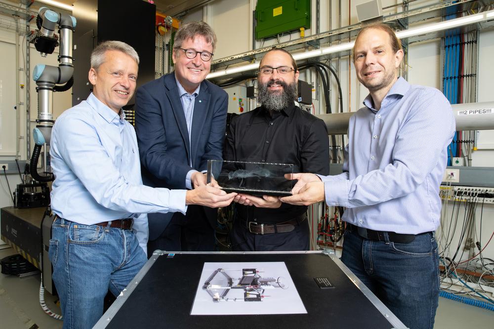
Large asteroid impacts can melt significant amounts of material from Earth’s crust (artist’s impression). Credit: NASA, Don Davis
Asteroid impacts are catastrophic events that create huge craters and sometimes melt parts of Earth’s bedrock.“ Nevertheless, craters are often difficult to detect on Earth, because erosion, weathering and plate tectonics cause them to disappear over millions of years,” Langenhorst explains. Therefore, minerals that undergo characteristic changes due to the force of the impact often serve as evidence of an impact. For example, quartz sand (which chemically is silicon dioxide, SiO2) is gradually transformed into glass by such an impact, with the quartz grains then being crisscrossed by microscopic lamellae. This structure can only be explored in detail under an electron microscope. It can be seen in material from the relatively recent and prominent Barringer crater in Arizona, USA, for example.
Read more on the DESY website
Image: Large asteroid impacts can melt significant amounts of material from Earth’s crust (artist’s impression)
Credit: NASA, Don Davis


